Consultant Laboratory for Diagnostic Electron Microscopy of Infectious Pathogens (Diagnostic EM of Pathogens)
Date: 04/09/2023
- Leader:
- Dr. Michael Laue
Diagnostic electron microscopy provides a rapid survey about pathogens present in a sample. All micro and nanoparticles, including the smallest known virus particles, can be detected based on morphological criteria, which allows to reveal and to describe even emerging pathogens. At best, the investigation by electron microscopy provides a quick tentative diagnosis which guides and speeds up a further, more specific diagnosis, or supports a symptomatic diagnosis. Additionally, diagnostic electron microscopy may exclude the presence of a suspected pathogen and serve as an independent control for the diagnosis by other methods.

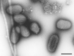
Parapoxviruses
Tasks
- Diagnostic electron microscopy of pathogens in clinical samples, cell cultures and environmental samples.
- Consulting of scientists, medical doctors and technical staff regarding diagnostic electron microscopy (e.g. choice of methods, second opinion in diagnostics).
- Advanced training in diagnostic electron microscopy (e.g. via lab courses) and support of quality assurance.
- Promotion of network activities in diagnostic electron microscopy, e.g. by conducting international workshops.
- Organisation of an external quality testing scheme for diagnostic electron microscopy of viruses.
If you need to send us samples for diagnosis, please contact us first before shipment to guarantee a proper sample collection and preservation for the shipment.
Projects
The Consultant Laboratory for Diagnostic EM is part of the „Advanced Light- and Electron Microscopy“ unit (ZBS 4) of the Robert Koch Institute. Apart from diagnostic electron microscopy, the unit performs ultrastructural and microbiological research on bacterial endospores and biofilm, and acts as a core facility for light and electron microscopy.
